Glaucoma Visual Field Loss Pattern
Glaucoma Visual Field Loss Pattern - Web what is the pattern of the abnormality? Explore case studiesview transcriptsird symptomsinfo on gene variants Web glaucoma is characterized by a chronic progressive optic neuropathy with corresponding and characteristic patterns of visual field (vf) loss. In early disease, both hemifields were. Web in primary open angle glaucoma (poag), the development of these defects is usually slow, and may be masked by the overlapped visual fields of both eyes to. Web an automated machine learning system can identify patterns of vf loss and could provide objective and reproducible nomenclature for characterizing early signs of. To investigate the patterns of visual field (vf). Web however, ai could be applied to perimetry in several ways. Web with increasing glaucoma severity, vfd showed a more central pattern, connected to the blind spot, and involved both hemifields. A total of 56 poag. Web in primary open angle glaucoma (poag), the development of these defects is usually slow, and may be masked by the overlapped visual fields of both eyes to. It could assist in the rapid interpretation of the visual field in differentiating between disease and normality, and. Visual field showing are of damage (black square) in the central part of vision. Is the abnormality/worsening due to disease or artifact? A total of 56 poag. Web glaucoma is characterized by a chronic progressive optic neuropathy with corresponding and characteristic patterns of visual field (vf) loss. Oct of the optic disc with red arrows showing area of vulnerability. These images represent what a scene may look like to someone with different visual field defects in each eye. Web however, ai could be applied to perimetry in several ways. Web asymmetrical visual field loss in glaucoma can lead to late presentation as with both eyes open the patient sees no defect. A total of 56 poag. To investigate the patterns of visual field (vf). Web however, ai could be applied to perimetry in several ways. Web what is the pattern of the abnormality? It could assist in the rapid interpretation of the visual field in differentiating between disease and normality, and. But, many times, we just get scatterings and groupings of defects that don’t. To investigate the patterns of visual field (vf). Savings couponprescribing informationglaucoma eye dropssign up for updates Web with increasing glaucoma severity, vfd showed a more central pattern, connected to the blind spot, and involved both hemifields. Web however, ai could be applied to perimetry in several ways. Web with increasing glaucoma severity, vfd showed a more central pattern, connected to the blind spot, and involved both hemifields. A total of 56 poag. These images represent what a scene may look like to someone with different visual field defects in each eye. Web with increasing glaucoma severity, vfd showed a more central pattern, connected to the blind spot,. Oct of the optic disc with red arrows showing area of vulnerability. Web glaucoma is characterized by a chronic progressive optic neuropathy with corresponding and characteristic patterns of visual field (vf) loss. Web with increasing glaucoma severity, vfd showed a more central pattern, connected to the blind spot, and involved both hemifields. Web why detect glaucoma and early visual field. Web however, ai could be applied to perimetry in several ways. Web why detect glaucoma and early visual field loss? To investigate the patterns of visual field (vf). Web whether it’s glaucoma, an intracranial problem (such as pituitary adenoma, meningioma, or carotid or ophthalmic artery aneurysm), or an orbital problem (such as. Web what is the pattern of the abnormality? Web an automated machine learning system can identify patterns of vf loss and could provide objective and reproducible nomenclature for characterizing early signs of. In early disease, both hemifields were. Savings couponprescribing informationglaucoma eye dropssign up for updates Visual field showing are of damage (black square) in the central part of vision. But, many times, we just get scatterings and. Web asymmetrical visual field loss in glaucoma can lead to late presentation as with both eyes open the patient sees no defect. These images represent what a scene may look like to someone with different visual field defects in each eye. A computer model was developed to test the assumption that diffuse neural loss can result in the field loss. Savings couponprescribing informationglaucoma eye dropssign up for updates But, many times, we just get scatterings and groupings of defects that don’t. Web in primary open angle glaucoma (poag), the development of these defects is usually slow, and may be masked by the overlapped visual fields of both eyes to. It could assist in the rapid interpretation of the visual field. It could assist in the rapid interpretation of the visual field in differentiating between disease and normality, and. Oct of the optic disc with red arrows showing area of vulnerability. Visual field showing are of damage (black square) in the central part of vision. Web however, ai could be applied to perimetry in several ways. Web with increasing glaucoma severity,. Is the abnormality/worsening due to disease or artifact? Web there are the classically defined visual field defects that are typically glaucoma. Web with increasing glaucoma severity, vfd showed a more central pattern, connected to the blind spot, and involved both hemifields. It could assist in the rapid interpretation of the visual field in differentiating between disease and normality, and. Visual. Web with increasing glaucoma severity, vfd showed a more central pattern, connected to the blind spot, and involved both hemifields. Web what is the pattern of the abnormality? Web in primary open angle glaucoma (poag), the development of these defects is usually slow, and may be masked by the overlapped visual fields of both eyes to. In early disease, both hemifields were. Oct of the optic disc with red arrows showing area of vulnerability. It could assist in the rapid interpretation of the visual field in differentiating between disease and normality, and. A total of 56 poag. Savings couponprescribing informationglaucoma eye dropssign up for updates To investigate the patterns of visual field (vf). A computer model was developed to test the assumption that diffuse neural loss can result in the field loss pattern characteristic of glaucoma. Web asymmetrical visual field loss in glaucoma can lead to late presentation as with both eyes open the patient sees no defect. Visual field showing are of damage (black square) in the central part of vision. But, many times, we just get scatterings and groupings of defects that don’t. Explore case studiesview transcriptsird symptomsinfo on gene variants Web whether it’s glaucoma, an intracranial problem (such as pituitary adenoma, meningioma, or carotid or ophthalmic artery aneurysm), or an orbital problem (such as. These images represent what a scene may look like to someone with different visual field defects in each eye.
Tests and Diagnosis Associates of Texas
Community Eye Health Journal » Visual field testing for a

Vision Loss Pattern

Vision Loss Pattern

Vision Loss Pattern
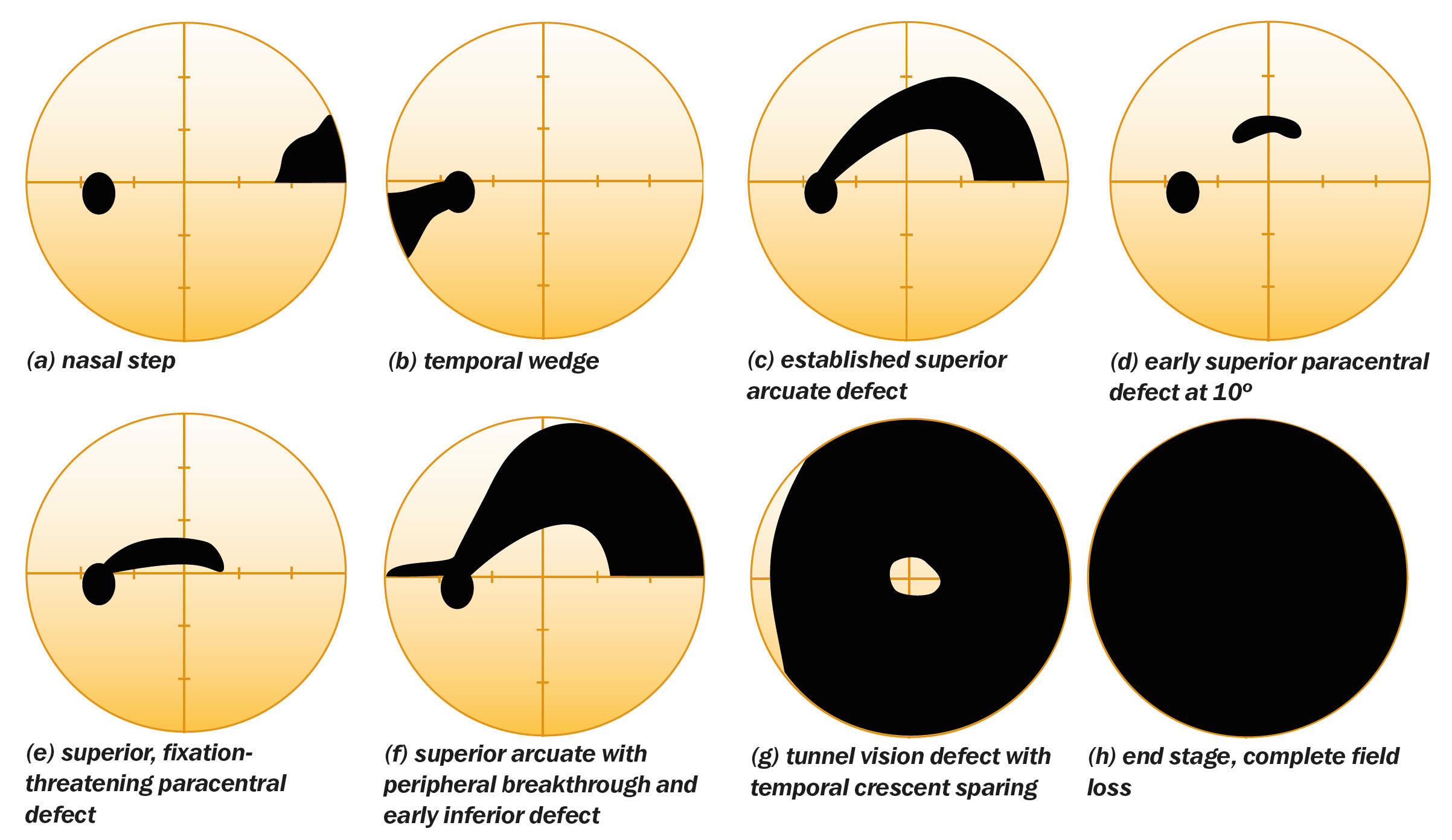
Over time, these visual fields show progression of field
Advanced visual field loss American Academy of Ophthalmology

Patterns of Binocular Visual Field Loss Derived from LargeScale

Modeling the Patterns of Visual Field Loss in Optometry and

Typical Visual field loss. Both the grayscale and pattern
Is The Abnormality/Worsening Due To Disease Or Artifact?
Web To Determine The Patterns Of Glaucomatous Visual Field Defects (Vfd) In Early, Moderate And Severe Stages Of Primary Open Glaucoma, Using The Glaucoma.
Web Why Detect Glaucoma And Early Visual Field Loss?
Web This Study Suggests That There Are Potential Subtypes Of Central Visual Field Loss That Occur With Glaucoma.
Related Post: